Recruiting the Lung While Looking at It!
Have you ever had a patient with a problematic atelectatic lobe that was very difficult to recruit? Understandably, you may have been afraid of using higher pressures because of over distending and potentially causing air leak. But what if you could have visualized the lung in real time during recruitment maneuvers?
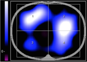
Electrical Impedance Tomography (EIT) may provide that window you were looking for. We came across this exact scenario in an eleven-year old girl with hypoxemic respiratory failure secondary to influenza A pneumonia. With EIT, (Dräger Pulmovista 500, Drägerwerk AG & Co. KGaA, Lübeck, Germany) even at a PEEP of 15 cm H2O, we could not open her right dorsal quadrant (a).
(a)
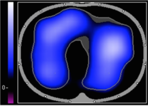
PEEP 22 cm H2O
PEEP 15 cm H2O
(b)
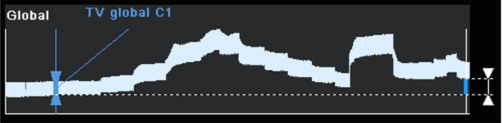
ΔEELI 30 minute trend
(c)
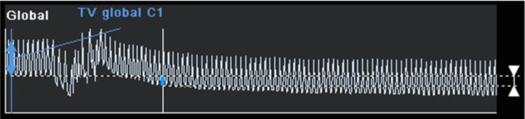
ΔEELI 5 minute trend
After an incremental/decremental PEEP maneuver (b), we found significant derecruitment and alveolar instability at PEEP < 22 cm H2O (c). After another recruitment and a final PEEP of 22 cm H2O, the improvements were obvious (a). Surprisingly, despite a significant increase of PEEP, her PIP dropped from 38 to 29 cm H2O with identical tidal volumes (5 ml/kg). During this maneuver, no air leak or hemodynamic instability was noted.
We reported this case (Dmytrowich et al., J Clin Monit Comput, 2018) to illustrate that EIT may “herald a new revolution in mechanical ventilation”. The benefits of real time images and ongoing data provide important information for safe recruitment strategies, alveolar stability and regional overdistention. Never before would we have pushed PEEPs so high, but EIT offered a window into the lungs that made it possible.
Robots in the North
There is a classic Star Wars scene when C-3PO tells R2-D2 after his long slumber, “Oh, my dear friend. How I’ve missed you”. This is…